Art-labeling activity the structure of a skeletal muscle fiber – Embark on an art-labeling expedition into the intricate world of skeletal muscle fibers, where each stroke of the brush reveals the fundamental building blocks of movement. Delve into the depths of this interactive learning experience, meticulously labeling the key components of these microscopic powerhouses, gaining an unparalleled understanding of their structure and function.
As you meticulously label each element, you’ll uncover the secrets of the sarcolemma, the protective sheath that envelops the muscle fiber. Journey into the depths of the sarcoplasm, the gel-like substance that houses the myofibrils, the thread-like structures responsible for muscle contraction.
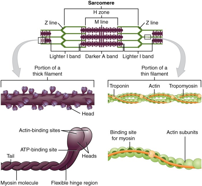
Explore the arrangement of myofilaments within a sarcomere, the basic unit of muscle contraction, and witness the interplay of actin and myosin, the molecular players that drive muscle movement.
Anatomy of a Skeletal Muscle Fiber: Art-labeling Activity The Structure Of A Skeletal Muscle Fiber

Skeletal muscle fibers are the basic units of muscle tissue. They are long, cylindrical cells that are responsible for generating force and movement.
The basic structure of a skeletal muscle fiber includes:
- Sarcolemma:The cell membrane of the muscle fiber.
- Sarcoplasm:The cytoplasm of the muscle fiber.
- Myofibrils:The contractile units of the muscle fiber.
- Myofilaments:The protein filaments that make up the myofibrils.
The myofilaments are arranged in a repeating pattern called a sarcomere. The sarcomere is the basic unit of muscle contraction.
The transverse tubules (T-tubules) are invaginations of the sarcolemma that run perpendicular to the myofibrils. The T-tubules allow the action potential to spread throughout the muscle fiber, triggering muscle contraction.
The sarcoplasmic reticulum (SR) is a network of tubules that surrounds the myofibrils. The SR stores calcium ions, which are released during muscle contraction.
Molecular Components of Skeletal Muscle Fibers
The major proteins involved in muscle contraction are actin, myosin, troponin, and tropomyosin.
Actinis a thin filament that forms the core of the myofibril.
Myosinis a thick filament that interacts with actin to generate force.
Troponinis a complex of three proteins that binds to actin and regulates muscle contraction.
Tropomyosinis a protein that binds to actin and prevents myosin from interacting with it.
The sliding filament theory of muscle contraction states that muscle contraction occurs when the myosin filaments slide past the actin filaments, causing the sarcomere to shorten.
Energy Metabolism in Skeletal Muscle Fibers, Art-labeling activity the structure of a skeletal muscle fiber
Skeletal muscle fibers use three main energy sources: ATP, creatine phosphate, and glycogen.
ATPis the primary energy source for muscle contraction.
Creatine phosphateis a high-energy compound that can be used to rapidly regenerate ATP.
Glycogenis a complex carbohydrate that can be broken down into glucose to produce ATP.
The metabolic pathways involved in energy production include glycolysis, oxidative phosphorylation, and the Krebs cycle.
Muscle fatigue occurs when the muscle fiber can no longer generate enough force to perform a given task. Recovery from muscle fatigue requires the restoration of ATP levels.
Control of Skeletal Muscle Contraction
The nervous system initiates and regulates muscle contraction.
The neuromuscular junction is the synapse between a motor neuron and a muscle fiber.
The different types of muscle contractions include isometric, isotonic, and eccentric contractions.
Isometric contractionsare contractions in which the muscle length does not change.
Isotonic contractionsare contractions in which the muscle length changes.
Eccentric contractionsare contractions in which the muscle lengthens while it is under tension.
Skeletal Muscle Fiber Types
There are three main types of skeletal muscle fibers: slow-twitch, fast-twitch, and intermediate fibers.
Slow-twitch fibersare slow to contract and relax, but they are resistant to fatigue.
Fast-twitch fibersare fast to contract and relax, but they fatigue quickly.
Intermediate fibershave properties that are intermediate between slow-twitch and fast-twitch fibers.
The muscle fiber type composition of a muscle is determined by genetic factors and training.
Adaptations of Skeletal Muscle Fibers to Exercise
Skeletal muscle fibers adapt to exercise training by increasing in size and strength.
These adaptations improve muscle strength, power, and endurance.
Nutrition and recovery play an important role in muscle adaptation.
FAQ Guide
What is the role of the sarcolemma?
The sarcolemma is the outermost layer of the muscle fiber, responsible for maintaining the integrity of the cell and regulating the exchange of substances between the muscle fiber and its surroundings.
What are myofibrils?
Myofibrils are elongated, thread-like structures within the muscle fiber that contain the myofilaments, the proteins responsible for muscle contraction.
What is a sarcomere?
A sarcomere is the repeating unit of a myofibril, representing the basic unit of muscle contraction.

